
The Anatomy Lab Heart Dissection form provides a comprehensive overview of the educational resources and procedures involved in studying the heart, a vital organ responsible for sustaining life through its relentless pumping action. This form notably outlines various stages of dissection, including external anatomy identification, cardiac damage assessment, and testing blood flow through a repaired heart, helping students to gain hands-on experience that reflects the practices of a cardiac surgeon. Covering key concepts in physiology, anatomy, and cardiovascular health, the form is designed for a wide range of learners, making connections between theoretical knowledge and practical application. It also details essential materials, prerequisites for preparatory activities, and the necessary tools for conducting dissections safely and effectively. Furthermore, recent research highlighted in the form emphasizes advancements in understanding cardiovascular diseases— a leading cause of mortality— and explores innovative treatments such as biomaterials and artificial blood. By engaging with this form, educators can facilitate a rich learning experience that not only nurtures students' understanding of heart anatomy but also raises awareness of the importance of cardiovascular health and advancements in medical science.
bbsrc.ac.uk
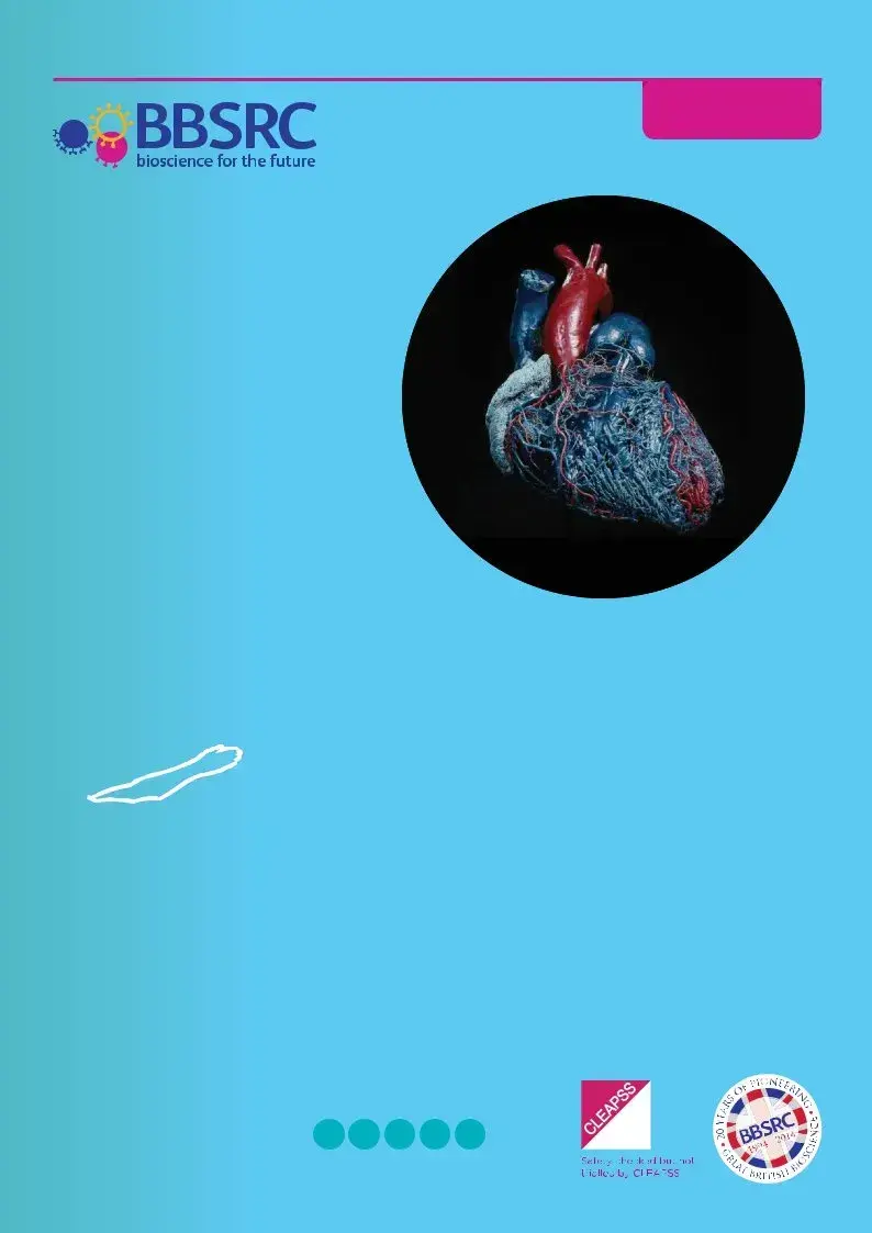
Heart Surgery
and Dissection
The heart is an amazing organ that continues to beat roughly every second, every day, for your entire life. That is 100,000 beats each day, and every minute about five litres of blood are pumped out of the heart. It has to keep working
Suitable for Key Stage:
1 2 3 4 5

Key Information |
Teacher |
|
Contents
02Key information
05Recent research
14Teacher preparation
15Health and safety
16Stage 1 – External anatomy
18Stage 2 – Identification and repair of heart damage
19Stage 3 – Testing blood flow through a repaired heart
20Stage 4 – Examining the internal anatomy of a heart
23Curriculum links
24Further reading
25How the heart works
29Stage 1 – External anatomy
30Stage 2 – Identification and repair of heart damage
31Stage 3 – Testing blood flow through a repaired heart
32Stage 4 – Examining the internal anatomy of a heart
33Autopsy report form
34Heart anatomy
35Card flow
36Circulation worksheet
37Missing Words
39Wordsearch
40Crossword
41Answers
44Glossary
View online
Scan the QR Code.
www.bbsrc.ac.uk |
22of4611 |
|
Key Information |
Teacher |
|
Science topics
Physiology, anatomy, cardiovascular system, exchange and transport, pathology, disease and injury
Resources
Age |
|
• |
Student sheets |
|
|
• |
PowerPoint presentation |
|
|||
|
|
|
|
|
|
|
|
Duration
125 minutes
Keywords
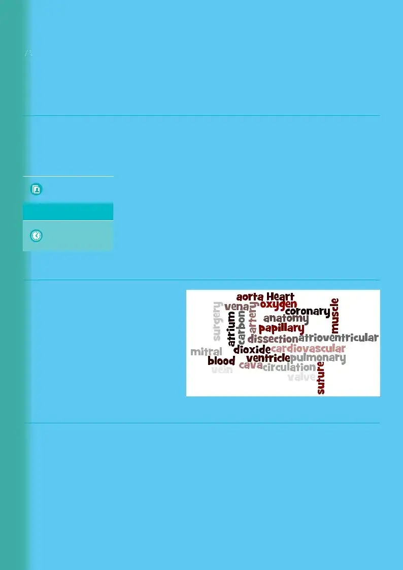
Heart, blood, circulation, cardiovascular, anatomy, dissection, surgery, ventricle, aorta, atrium, muscle, valve, coronary, pulmonary, artery, vein, vena cava, suture, papillary, atrioventricular, mitral, oxygen,
carbon dioxide
Learning outcomes
Students will be able to:
•Identify the internal and external anatomy of a heart
•Dissect a heart and be able to model the techniques of a heart surgeon
•Discuss heart diseases and disorders, describe how they occur, and name risk factors and possible preventative measures.
www.bbsrc.ac.uk |
32of4611 |
|

Key Information |
Teacher |
|
What you will need
•Sheep or lamb hearts
•Rubber tubing and syringe
•Dissecting equipment – trays, pins or cocktail sticks, forceps, blunt probes,
•Masking tape or stickers
•Marker pens
•Washing up bowls or access to sinks
•Curved needles
•Dental floss or fishing line
•Rulers
•Eye protection
•Waterproof aprons
•Balance
Optional
•Disposable nitrile or vinyl gloves
•Scalpels
•Slides featuring heart muscle tissue and cardiovascular pathology
•Camera for students to record the progress of their activity
Prior Learning
Students should carry out a preparatory activity to familiarise themselves with the structures of the heart. Resources such as worksheets, animations and videos can ensure students get the most from the learning session. A description of how the heart works and diagrams of the heart and circulatory system for students to label are provided.
|
Equipment |
|
|
|
|
|
© BBSRC |
|
|
|
|
www.bbsrc.ac.uk |
|
42of4611 |
|
|
|
Recent Research
The Biotechnology and Biological Sciences Research Council (BBSRC) is working towards lifelong health and
Key issues include linking changes at the molecular and cellular level to those observed at the tissue and whole organism level. A large body of evidence demonstrates that the quality and quantity of food, and dietary choice affects ageing and lifespan. There are also good data that aerobic exercise increases healthy lifespan, improves regulation of glucose metabolism and can reduce
From stem cells to artificial blood, researchers are investigating treatments for broken hearts and a variety of cardiovascular diseases. Work on biomaterials and tissue engineering is producing improved medical implants and devices such as pacemakers, as well as a range of substances with
Throughout this research, BBSRC encourages work that adopts the principles of the 3Rs (Replacement, Refinement and Reduction) in the use of animals, and aims to improve animal welfare.
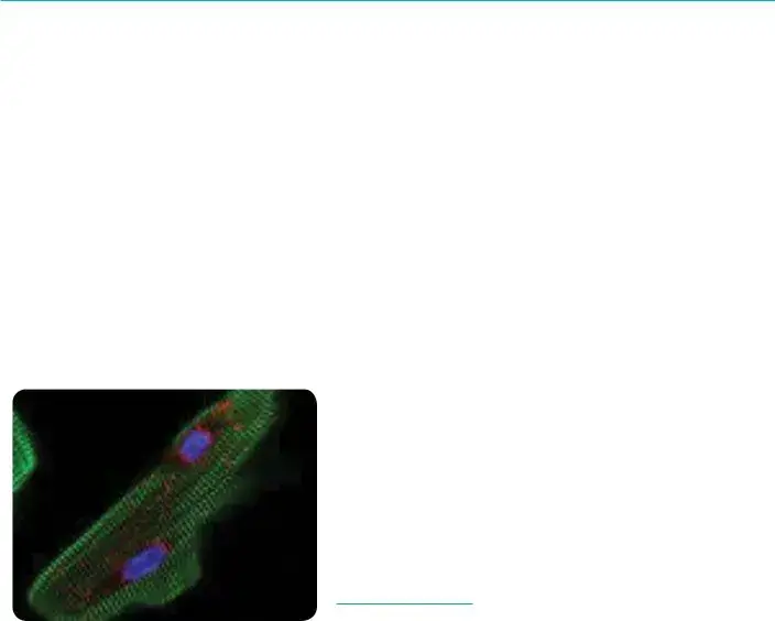
Change at cellular level
© Babraham Institute
Recent Research
Can we ever mend a broken heart
Scientists at the University of Nottingham are working towards a treatment for damaged hearts. Heart disease is the most common cause of death in the UK and each year there are 20 million cases around the world. About one in five men and one in eight women die of heart disease but in the future we might be able to mend broken hearts with new heart cells.
Our hearts can fail because they are getting old, because of the stresses and strains placed on our heart by the drugs we take to treat illness such as cancer, or simply because of our genetic
Scientists are trying to develop techniques that would turn some of our own skin cells into stem cells and then turn these into beating heart cells to replace lost or damaged cells. There would be no need for drugs and we could be healed with a simple injection of our own cells.
Developing techniques
© Luchschen

Recent Research
The answer to high blood pressure may be in our brains
Blood pressure is controlled by our brains and our kidneys. Scientists are now beginning to look more closely at the brain and genes involved in the development of high blood pressure. Having high blood pressure, known as hypertension, increases the risk of stroke, heart attacks and kidney failure. You can’t ‘feel’ whether you have high blood pressure, which makes it so dangerous. Nearly one billion people around the world have hypertension.
The kidney controls blood pressure by regulating the amount of water and salt reabsorbed into the blood. High levels of salt in the blood cause the kidneys to retain water and lead to raised blood pressure. Most high blood pressure treatments target the kidneys but new research will look at ways to target the nervous system. The brain detects blood pressure using the carotid sinus, a small swelling in the carotid artery. The carotid sinus has stretch receptors that send signals to the brain when blood pressure rises. These signals go to the cardiovascular centre in the medulla and a negative feedback system sends out signals to lower heart rate and dilate blood vessels to lower the pressure.
For up to 50 per cent of patients on blood pressure tablets the treatment is ineffective and many suffer from unpleasant side effects. Researchers will explore how the genes in the brain trigger hypertension and study how ageing, exercise and a condition known as sleep apnoea affect the activity of these genes. Sleep apnoea is a blockage of airways during the night that cuts off oxygen and causes people to wake. It is associated with obesity and is often accompanied by loud snoring.
To find novel drug targets and improve current treatments, scientists will use tissue from brain banks to determine how genes are regulated in specific brain regions and how these genes interact during the development of high blood pressure. The scientists will also study how the environment may affect gene activity. In order to do this they will use a special technique that allows them to record the activity of single nerve fibres that control the diameter of arteries.
Recent Research
What causes a big heart?
Under some conditions heart cells get larger in a process known as hypertrophy. While this may sound romantic, and is necessary for developmental growth, hypertrophy often leads to heart failure. If there is a
Conditions like high blood pressure also cause the heart to grow, but in this case increased size does not improve the heart’s pumping capacity. Instead it promotes the transition to cell death and heart failure as the heart becomes prone to irregular heartbeats – arrhythmias. Researchers have discovered how signalling processes within the heart can trigger the development of enlarged heart cells which lead to heart failure. The discovery provides new insight into the mechanisms controlling cardiac growth and the processes that cause adaptation and remodelling of heart muscle. Cardiac failure accounts for 25% of deaths in the UK and is a primary cause of death in the elderly. Understanding how these pathological changes occur in the heart, in response to disease and ageing, may reveal therapeutic targets and new approaches to the treatment of heart disease.
The research team found that a tiny molecule made of ribonucleic acid (RNA), microRNA, controls the levels of specific receptors produced in heart cells. MicroRNAs are copied from deoxyribonucleic acid (DNA) but do not contain code for protein. Rather, they control gene activity by binding
to specific related sequences. It is the interactions between these microRNA molecules and the receptors that promote hypertrophic remodelling of heart muscle. The receptors are channels controlling the movement of calcium ions, which are an important ‘messenger’ inside cells, regulating heart rhythm and function. Calcium ions are the link between electrical excitation of a muscle cell and its contraction. When an electrical impulse arrives at a muscle it causes calcium to enter the cell and releases calcium from internal stores resulting in contraction of the cell. If calcium signals occur at the wrong place or time, for example due to changes in receptor regulation, this can change the heart structure – decreasing its ability to pump efficiently, or triggering irregular heartbeats.
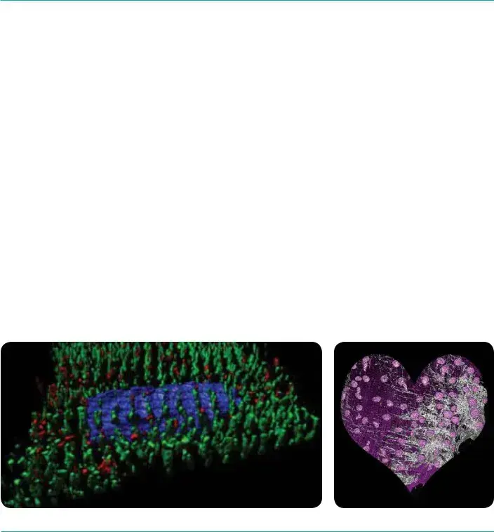
3D Reconstruction of a section through a rats heart
© Dr Llewelyn Roderick Group

Recent Research
Scientists discover the cause of a broken heart
Around
Researchers now think this ‘broken heart syndrome’ is a protective response to very high levels of adrenaline released during stress. Instead of stimulating the heart, the body responds to the adrenaline by reducing its pumping power. The same condition is sometimes seen in people who are injected with adrenaline to treat severe allergic reactions. Therefore drugs that stimulate adrenaline are likely to make the condition worse. The scientists used their animal model of the disease to investigate suitable treatments and found beneficial drugs that stimulate the heart using a different pathway to adrenaline.
Recent Research
Why some people endure exercise better than others
Exercise is essential for maintaining good health. An understanding of how elite athletes’ bodies function may help prevent elderly people developing chronic illnesses.
Researchers are studying elite athletes using
Computer models will be used to improve our understanding of why exercise tolerance is limited in sedentary or elderly individuals. The findings will be used as the basis for treating patients with heart and lung conditions who have problems with exercising.
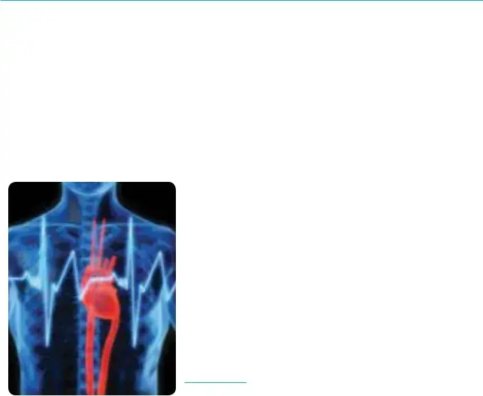
Human Heart
© Thinkstock
| Fact Name | Description |
|---|---|
| Heart Function | The heart beats approximately 100,000 times daily, pumping about five liters of blood per minute. |
| Scientific Research | Researchers utilize mathematics and imaging techniques to enhance understanding of heart function and disease. |
| Age Group | This dissection form is suitable for students aged 14 to 18 years, aligning with educational standards. |
| Learning Outcomes | Students will learn to dissect a heart, identify anatomical features, and discuss heart diseases and their prevention. |
| Key Equipment | Required tools include dissecting equipment, rubber tubing, and protective gear like goggles and aprons. |
| Subject Areas | This lab covers topics such as physiology, cardiovascular health, and surgical techniques. |
| Governing Laws | Research and dissections must comply with ethical standards set forth by the Biotechnology and Biological Sciences Research Council (BBSRC). |
As you prepare to fill out the Anatomy Lab Heart Dissection form, it is essential to follow each step meticulously. This ensures that the necessary information is captured accurately, enhancing the overall learning experience for all participants involved in this fascinating exploration of the heart.
The Anatomy Lab Heart Dissection form is a resource designed for educational purposes, specifically for students aged 14 to 18. It outlines various activities related to heart anatomy, dissection, and surgery techniques. The resources provided help students understand the structure and function of the heart, as well as common cardiovascular diseases and their risk factors.
To successfully conduct the heart dissection, you will need the following materials:
Optional items include disposable gloves, scalpels, slides with heart muscle tissue, and a camera to document the dissection process.
Students can achieve several learning outcomes through the heart dissection activities, such as:
This practical experience enriches their understanding of human physiology and anatomy.
Yes, prior preparation is highly encouraged. Students should engage in activities that will help them familiarize themselves with the structures of the heart. This might include reviewing worksheets, animations, or videos. Additionally, providing diagrams for students to label can enhance their comprehension of the heart and circulatory system.
Recent research focuses on a variety of areas to improve heart health. Scientists are investigating:
Such research plays a crucial role in understanding and potentially preventing heart disease, which is a leading cause of death.
Maintaining safety during the dissection is crucial. Here are some key safety measures:
By following proper safety protocols, students can focus on learning while minimizing risks.
Filling out the Anatomy Lab Heart Dissection form can be a straightforward process, but many people make common mistakes that can compromise their understanding and results. Here are seven mistakes to be aware of while completing the form.
First, many forget to review the instructions thoroughly before beginning. This oversight can lead to skipping necessary information or misinterpreting the questions. Attention to detail is crucial, particularly in a scientific context where accuracy matters.
Another frequent mistake occurs with **spelling errors** in anatomical terms. These errors can cause confusion or miscommunication when working with peers or instructors. Double-checking spelling, especially for technical words like "ventricle" or "atrium," is essential.
In addition, some individuals may neglect to properly label diagrams required on the form. Diagrams should be clear and well-labeled, as they are often a critical part of demonstrating understanding. Failing to meet this requirement can affect the overall clarity of the work.
Moreover, people often rush through the identification of risk factors associated with heart diseases and disorders. A shallow analysis can lead to incomplete responses. Taking the time to think through and research these factors can enhance the quality of the submission.
Some individuals also forget to include relevant research citations or references. Supporting your statements or findings with credible sources establishes reliability and allows others to follow your thought process. Leaving this out can weaken the overall impact of the work.
Lastly, many fail to reflect on personal learning outcomes and experiences from the dissection. This reflection is not only beneficial for personal understanding but allows for richer discussions with peers and teachers. Ignoring this part of the task can lead to missed opportunities for improvement.
By avoiding these common mistakes, participants can ensure their completion of the Anatomy Lab Heart Dissection form is not only accurate, but also educational. Act promptly to correct any oversights and enhance your understanding of the intricacies of heart anatomy and function.
In addition to the Anatomy Lab Heart Dissection form, several other documents and forms are typically utilized in this context. Each of these documents serves a specific purpose, facilitating the organization and clarity of the dissection process and enhancing the learning experience for students. Below is a brief overview of five key documents that may accompany the dissection activity.
These documents collectively enhance the educational value of the heart dissection experience, ensuring that students are well-prepared and informed as they explore the intricate workings of this vital organ. Together, they foster a deeper understanding of human physiology and promote a thorough examination of heart health.
When filling out the Anatomy Lab Heart Dissection form, consider the following guidelines to ensure a smooth process:
Understanding the Anatomy Lab Heart Dissection form is essential for effective dissection and learning. However, several misconceptions may hinder the process. Here are 8 common misconceptions:
Understanding how to effectively use the Anatomy Lab Heart Dissection form is essential for a successful learning experience. Here are some key takeaways to consider: